USA-Made Hyperbaric Chambers Ship within 2-3 days
Talk to an Expert
512.789.2788
December 4, 2023
According to the CDC, drowning is a leading cause of death in children aged 1 to 14. It is extremely dangerous as it deprives the entire brain of necessary oxygen. Often, this deprivation leads to the death of brain cells or the death of the drowning victim. In fact, brain cells begin to die within five minutes of oxygen deprivation. The parts of the brain that are most often affected are the parts that control memory, speech, and movement. That is why survivors of drowning usually suffer from deficits in memory, speech, and movement. The brain injury they experience is called anoxic brain injury or ABI.
This type of brain injury involves a temporary loss of blood supply to the brain. This is the most devastating type of brain injury and is usually seen after drowning, cardiac arrest, or birth injury. With an anoxic brain injury, all areas of the brain can be affected. However, the combination and severity is affected by a few factors.
Anoxic brain injuries can have a variety of symptoms based on the factors above. The effects will depend on which parts of the brain are affected, which are different from person to person. Some of the common symptoms are:
Many times, patients also experience seizures. Most patients have severe impairments and are often only minimally conscious, making them totally dependent.
Healing of the brain – or any part of the body – cannot take place if the tissue and cells do not receive the appropriate oxygen levels. The benefits of hyperbaric oxygen treatment for drowning are maximal when the treatment is delivered close to the time of the anoxic injury. While this is the case for all medical conditions, it is especially so for anoxic brain injuries. When delivered within three hours of the anoxic injury, HBOT can prevent as much as 90% or more of the injury, particularly the secondary more devastating component of injury which occurs when blood flow and oxygen are restored. Both the initial injury from loss of oxygen and the secondary injury when a heart beat is regained cause the loss of large amounts of both gray and white matter. The gray matter is the outer and deep parts of the brain containing brain cells and the white matter is the connecting tracts of the brain.
Below are Case Studies by Renown Hyperbaric Doctor, Dr. Harch
Below are two drowned children. The first is from Dr. Harch’s initial group of 41 drowned children treated from 1989-2016. All, but one, were at least one year after drowning. This child is a 4 year old boy who was treated with HBOT in 1993, two years post-drowning. He is wheelchair bound with global deficits in cognition, speech, motor, and coordination. The first SPECT brain blood flow scan is before his first HBOT, the second is 2 hours after his first HBOT, and the 3rd is after 80 HBOTs. At the time of the 3rd brain scan this little boy had experienced an improvement in spasticity, movement of all 4 extremities, increase in trunk and head control as well as improvements in swallowing, awareness, non-verbal communication, and attention span. An HBOT-generated global increase in blood flow on the 3rd scan was responsible for all of these improvements.

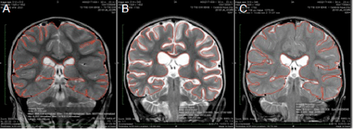
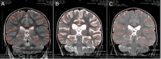
The second child is a two year old girl who was the first child in Dr. Harch’s second group of over 60 drowned children, nearly all of whom were treated within 6 months of drowning from 5/2016 to present. Her case was published in Medical Gas Research in 2017 (https://www.medgasres.com/temp/MedGasRes72144-6908434_191124.pdf). Her MRI scans below show a global loss of brain tissue one month after her drowning and a near complete regrowth and restitution of brain volume after normobaric oxygen by nasal cannula and hyperbaric oxygen. At the time of initiation of the nasal cannula oxygen she was a thrashing semi-conscious little girl. At the time of the final MRI she was walking and talking (see the videos in the link above).
The MRI slices below are what are called coronal slices, a view from the top of the head to the bottom. In each slice you are looking directly at this little girl. The left side of the image is the right side of her head. MRI scan A is 3 days after drowning. MRI scan B is 31 days after drowning where there has been a 12.3% loss of brain tissue, seen as increased white color on the scan (white is the cerebrospinal fluid or water of the brain). MRI scan C is 27 days after her 40th HBOT or 162 days after drowning. This is normally the time when one sees near-maximal loss of brain tissue. Instead, she has experienced a 13.9% increase in brain tissue.
https://www-ncbi-nlm-nih-gov.ezproxy.lsuhsc.edu/pmc/articles/PMC5510296/
Share on:
A free, open discussion group to explore hyperbaric oxygen therapy, compare home chamber brands, access expert info, and get exclusive member discounts.